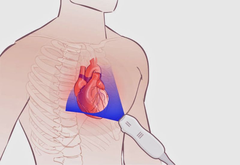
Cardiac ultrasound, also known as echocardiography, is an imaging method used to evaluate the heart’s structure, function, and potential abnormalities. Depending on the individual case, different echocardiographic techniques may be prescribed by healthcare professionals. This article explores the key details of this essential diagnostic tool.
1. Cardiac Ultrasound: A Crucial Imaging Tool in Heart Health Assessment
1.1 What is Cardiac Ultrasound?
The heart is a vital organ responsible for pumping blood to nourish cells, tissues, and organs throughout the body. Any abnormalities in its structure or function can significantly impact overall health.
To address this, diagnostic techniques for early detection and management of heart conditions are continually evolving. Among these, cardiac ultrasound remains a fundamental tool. Although not a new technology, it is often the first-line diagnostic test recommended when heart-related issues are suspected.
1.2 Common Cardiac Ultrasound Techniques
There are five primary types of echocardiography currently in widespread use:
– Transthoracic Echocardiography (TTE): A transducer is placed on the chest wall to generate images of the heart. A special gel is applied to facilitate the transmission of sound waves, providing clear visuals of the heart’s structure and function.
– Transesophageal Echocardiography (TEE): A smaller probe is guided through the throat into the esophagus, offering detailed images of the heart using sound waves.
– Doppler Ultrasound: This technique measures blood flow and pressure within the heart, aiding in the detection of pulmonary hypertension and other cardiovascular issues.
– 3D Echocardiography: This advanced method assesses heart valve function, reconstructs heart structures, and evaluates congenital heart defects in infants and children. It is also useful for pre- and post-surgical evaluations.
– Stress Echocardiography: Performed during physical exertion, such as running or walking on a treadmill, this test monitors heart rhythms and electrical activity to detect abnormalities under stress conditions.

An echocardiogram can help detect almost all heart-related issues.
2. Why is Cardiac Ultrasound Important?
Cardiac ultrasound allows physicians to identify heart problems with precision. By capturing real-time images of the beating heart, the technology helps determine how effectively the heart pumps blood, highlighting potential issues such as:
– Valve Disorders: For example, mitral valve regurgitation can be detected by observing the movement and function of heart valves.
– Blood Flow Analysis: It measures blood velocity through heart valves, essential for diagnosing conditions like aortic stenosis.
– Congenital Heart Defects: Identified during pregnancy or in newborns and young children.
– Heart Failure Monitoring: Evaluates the effectiveness of treatments for heart failure.
– Arrhythmias: Detects abnormal heart rhythms by assessing the heart’s motion and pinpointing underlying causes.
– Clots or Tumors: Accurately identifies the location of masses or blood clots within the heart.

Cardiac ultrasound aids in measuring the left ventricular ejection fraction to assess the effectiveness of heart failure treatment methods.
3. When Should Cardiac Ultrasound Be Performed?
Echocardiography is typically recommended for individuals experiencing potential heart-related symptoms, such as:
– Dizziness, lightheadedness, or headaches accompanied by chest pain or pressure.
– Irregular heart rhythms, such as rapid or slow beats, often causing shortness of breath.
– Vomiting, sudden breathlessness, or chest pain linked to heart injury.
– Difficulty engaging in physical activities due to a sensation of breathlessness or rapid heartbeats.
– Sudden vision dimming with nausea, potentially indicative of acute heart damage and circulatory issues.
– Other symptoms may include back, shoulder, neck, or arm pain, which are less commonly associated with heart disease but should not be ignored.

If you experience dizziness, lightheadedness, or chest pain, it is essential to visit a medical facility for evaluation.
Cardiac ultrasound is now widely available across healthcare facilities. However, the accuracy of results depends not only on advanced equipment but also on the expertise of medical professionals. At Thu Cuc TCI Healthcare System, patients benefit from modern technology and experienced cardiologists with over 30 years of diagnostic excellence. The compassionate, well-trained staff ensures a comfortable and professional experience.
This article aims to provide insights into cardiac ultrasound, helping patients better understand this essential diagnostic technique for heart health.


