Routine prenatal ultrasound is a cornerstone of prenatal care, offering invaluable insight into fetal well‑being and guiding pregnancy management. Understanding the appropriate regular prenatal ultrasound schedule ensures both peace of mind and medical vigilance. This article outlines the key routine prenatal ultrasound milestones, their medical significance, and practical preparation for each session.
1. What Is Routine Prenatal Ultrasound?
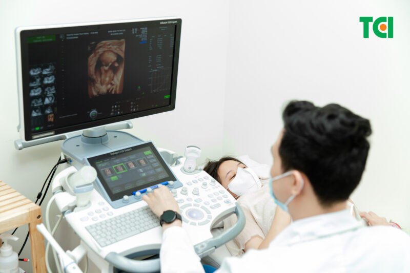
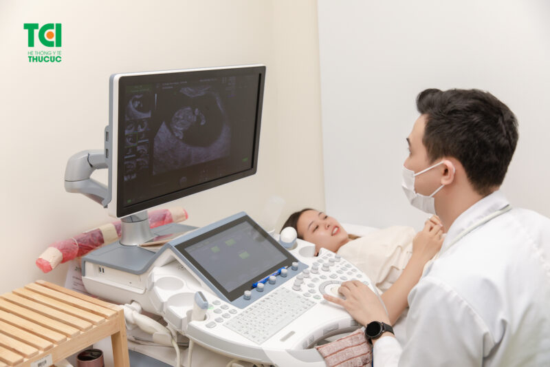
Routine prenatal ultrasound is a non-invasive imaging method that utilizes high-frequency sound waves to visualize the fetus and maternal reproductive organs. It is safe, painless, and essential for ongoing fetal assessment. These sessions typically align with prenatal checkups, ensuring timely data collection throughout pregnancy.
Ultrasound can be performed through two approaches:
– Transvaginal ultrasound: Utilizes a small probe inserted into the vagina, ideal for early pregnancy or when highly detailed images are needed.
– Transabdominal ultrasound: Involves moving a gel-covered probe over the abdomen; this is the standard practice for ongoing routine obstetric ultrasound sessions.

Prenatal ultrasound monitors the fetus using high-frequency sound waves to capture images from inside the abdomen.
At advanced stages, specialized techniques such as 2D, 3D, 4D, 5D, and Doppler ultrasound can further enhance the quality of fetal assessment.
2. Why Routine Prenatal Ultrasound Matters
Routine prenatal ultrasound fulfills several critical roles in pregnancy monitoring:
– Determining fetal age and estimated due date
– Observing fetal anatomy and developmental milestones
– Detecting congenital anomalies at various stages
– Measuring fetal growth and weight
– Evaluating placenta position, amniotic fluid levels, and maternal reproductive health
– Informing delivery planning and medical interventions
By obtaining consistent routine obstetric ultrasound data, physicians can offer timely advice tailored to each mother’s condition and pregnancy progression.
Regular prenatal ultrasound helps assess fetal health, detect abnormalities, and guide timely intervention.
3. Key Routine Prenatal Ultrasound Milestones
3.1 Early Pregnancy – Missed Period
Using regular prenatal ultrasound when a period is missed helps confirm intrauterine pregnancy. If no gestational sac is visible, blood tests may be used to verify pregnancy status.
3.2 Around Week 7
At this routine prenatal ultrasound stage, doctors assess fetal development, yolk sac, and fetal heartbeat—a reassuring sign of pregnancy viability.
3.3 Weeks 11–13
This is a crucial window for regular prenatal ultrasound involving nuchal translucency measurement and chromosomal screening. The scan can detect early markers of Down syndrome and other abnormalities. Gestational age, placental position, and number of fetuses are also evaluated.
3.4 Weeks 18–22
During the anatomy scan, routine prenatal ultrasound examines fetal organs, skeletal structure, and facial features. It can identify issues such as cleft lip, neural tube defects, and cardiac anomalies. Additional assessments include cervical length, placenta health, and amniotic fluid volume.
3.5 Weeks 24–28
This stage of routine prenatal ultrasound monitors fetal growth, positioning, and fluid balance. It also coincides with maternal gestational diabetes screening, helping refine nutritional and care plans.
3.6 Weeks 30–32
By this regular prenatal ultrasound milestone, fetal growth is substantial (up to ~1.7 kg), and anatomical development is nearly complete. The scan evaluates fetal position, organ function, and guides nutritional recommendations for late pregnancy.
3.7 Beyond Week 36
Weekly prenatal ultrasound sessions after week 36 track fetal heart rate, positioning, and amniotic fluid, ensuring timely intervention when delivery is near.
In addition to evaluating the fetus, ultrasound also screens for issues like amniotic fluid levels, placenta position, and uterine health.
4. What to Expect and How to Prepare
Each prenatal ultrasound typically takes about 30 minutes. Preparation tips include:
– Hydrate: Drink water and maintain a full bladder for transabdominal scans.
– Dress comfortably: Opt for clothing that allows relaxed access to the abdomen.
– Bring questions: Prepare queries about fetal development, results interpretation, or future care.
During the scan, a gel is applied, and the doctor uses a probe to examine the uterus. They explain the images and record key measurements. At session end, you’ll receive printed results and personalized advice on nutrition, testing, or follow-up care.
5. Guidelines and Best Practices
To maximize the effectiveness of routine obstetric ultrasound:
– Adhere strictly to the recommended scan schedule
– Choose a reputable healthcare provider with modern ultrasound technology
– Avoid unnecessary scans – follow medical guidance
Understand that while ultrasound is safe, it cannot detect every anomaly. Some conditions may be undetectable until after birth
6. Prenatal Ultrasound Services at Thu Cuc TCI
Thu Cuc TCI offers comprehensive maternal care packages incorporating routine obstetric ultrasound at all critical milestones. With advanced imaging systems and expert obstetricians, the facility ensures precise diagnostics and integrated care from early pregnancy through delivery. Their structured schedule, combined with reminders and professional interpretation, supports a worry-free pregnancy journey for international and domestic patients.
Routine prenatal ultrasound is indispensable in managing a healthy pregnancy. By understanding when and why each scan is performed, mothers gain confidence and maintain proactive engagement in their prenatal care. Choose a trusted provider like Thu Cuc TCI to ensure a well-supported and medically sound pregnancy experience.